The QUATTRO S environmental scanning electron microscope (FEG-ESEM) is equipped with a Dual EDS (60 m m2) / µ-XRF coupling.
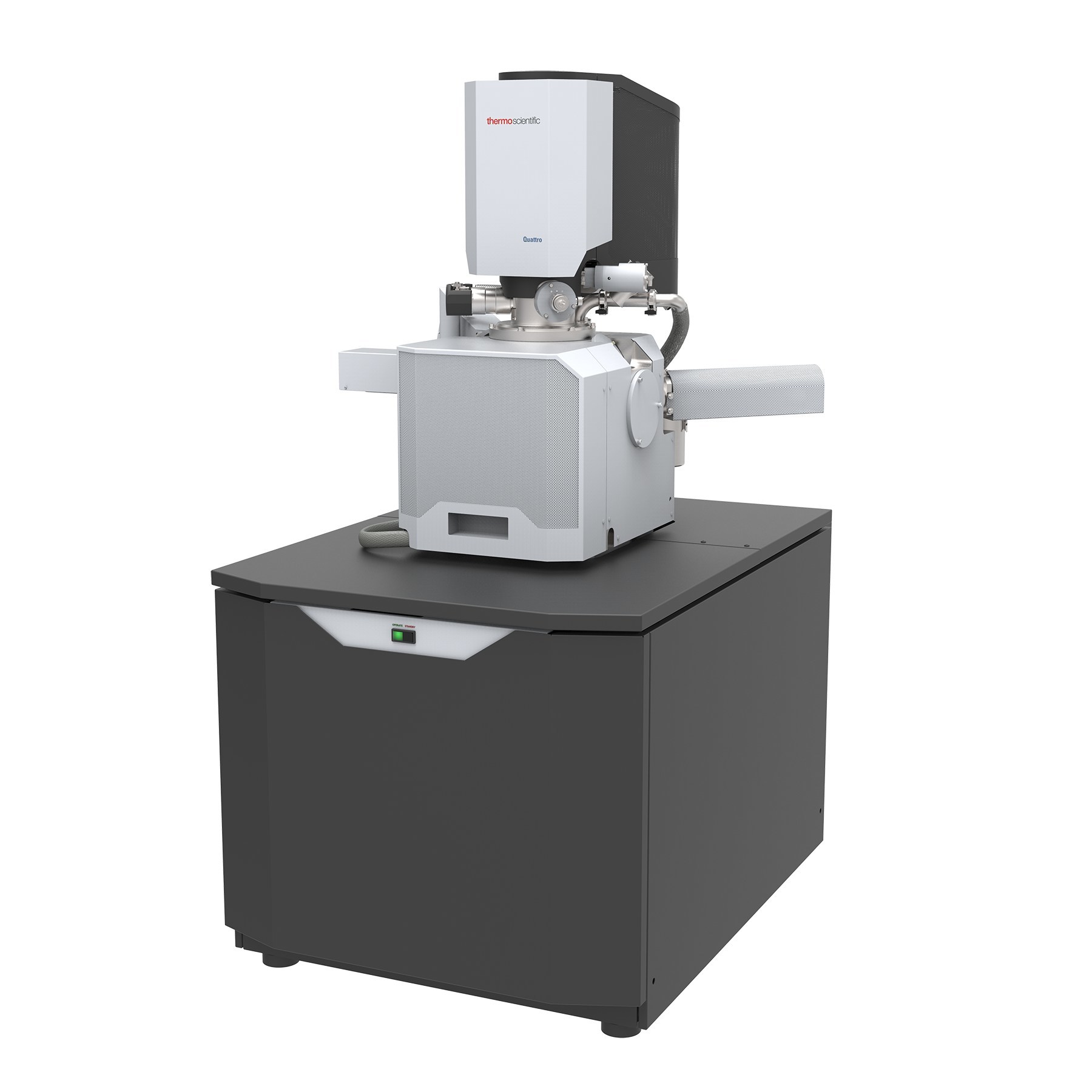
Special features:
- Modes: High Vacuum (<6*10-4 Pa), Gradient Vacuum (10 Pa to 130 Pa) , ESEM (10Pa to 4000 Pa).
- 5-axis motorized stage
- NavCam: 6 megapixels with digital zoom, co-located with the stage.
- Field emission cannon: voltages 200V-30 keV, current up to 200 nA.
- Resolution: 1.0 nm (high vacuum) to 1.3 nm (ESEM) in SE at 30 keV; 3.0 nm at 1 keV.
- Magnifications: x6 – x1,000,000
- Conventional Everhardt-Thornley high-vacuum secondary electron detector
- High-field gradient vacuum gas secondary electron detector.
- 4-segment diode backscattered electron detector
- IR camera
- 2x QUANTAX 200 EDS detector (60 mm2) in symmetry (Bruker)
- Canon X XTrace 400i (Bruker) with <35 µm capillary for µ-XRF
- RapidStage piezoelectric stage for µ-XRF mapping
- Licenses MAPS (correlative microscopy), TOPOMAPS (3D reconstructions), ESPRIT2 (EDS and XRF acquisition and processing)
Variable pressure:
In gradient vacuum or ESEM mode, samples can be observed without any preparation, whether they be insulating, hydrated or oily, at the cost of a slight compromise in spatial resolution in SE, BSE and EDS (there is no consequence for µ-XRF).
Nominal resolutions are achieved in high-vacuum mode, but they require the deposition of a conductive layer on the surface of insulating samples. The laboratory has AU/Pd and C metallizers for this purpose.
Electron imaging:
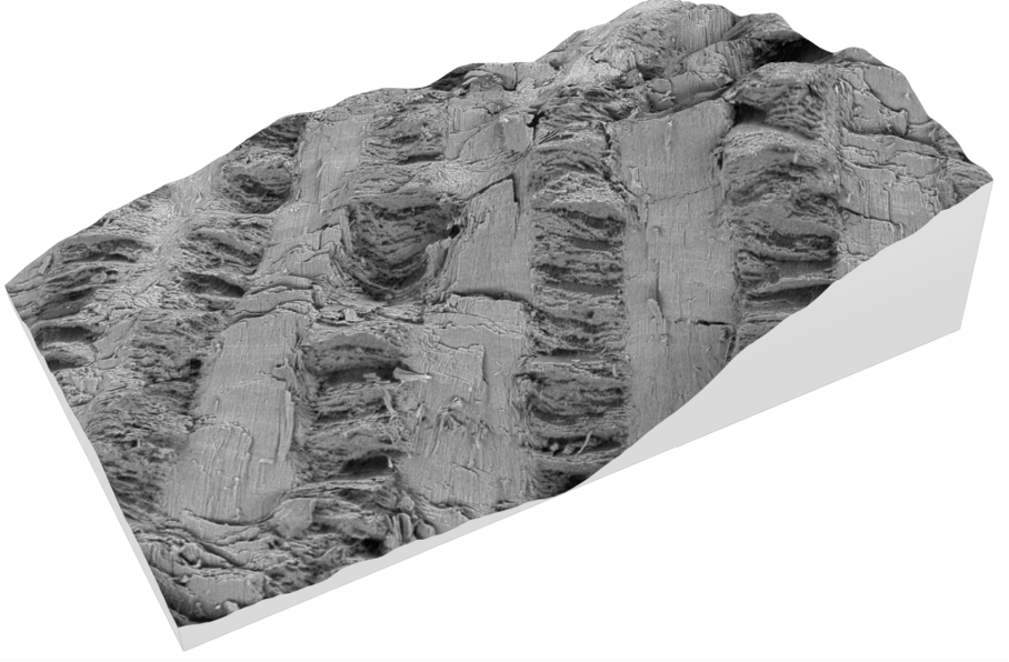
Secondary electron (SE) detectors provide topography contrast, enabling surface details to be imaged, even at very low accelerating voltages. The segmented diode backscattered electron (BSE) detector is primarily sensitive to atomic weight contrast, and can thus reconstruct a chemical contrast image of the sample at 2 keV and above.
The segmented BSE detector is compatible with TopoMaps Advanced software, and enables 3D reconstruction based on the three outer segments acquired in angular configuration (ABS), making it possible to restitute surfaces in 3D, calculate distances and slopes, extract object contours, etc.
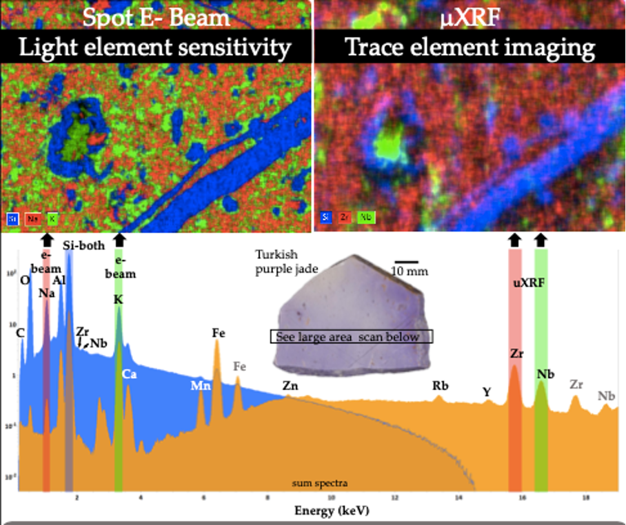
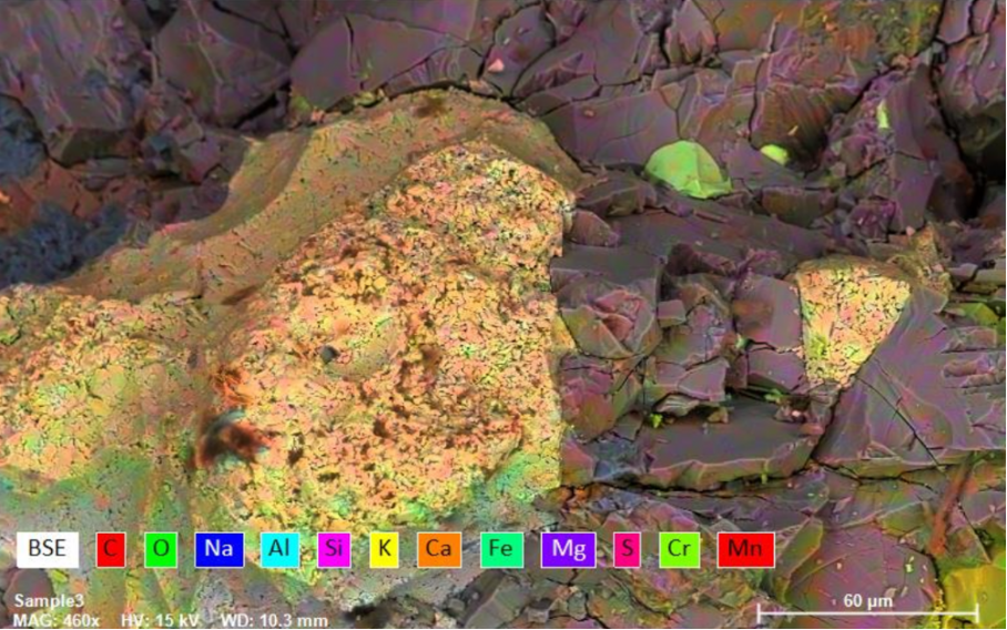
Energy dispersive X-ray spectroscopy:
The QUANTAX microanalysis system (Bruker) comprises two XFlash detectors with an active area of 60 mm2, and achieves an output count rate of 600 kcps per detector (at 66% dead time), i.e. 1.2 Mcps for both detectors. This system is therefore particularly well-suited to fast, quantitative elemental analysis, including mapping, with detection thresholds > 1000 ppm and spot sizes of the order of 1-2 µm at 15 keV.
X-ray micro-fluorescence:
The Quantax µ-XRF system consists of an X-ray gun (50 keV), and uses both XFlash detectors, benefitting from its fast acquisition times. Easy to set up (i.e. for insulating samples), it is suitable for precise quantification, including trace elements (detection thresholds ~50ppm for many elements), but with less spatial resolution than EDS (of the order of 40 µm). The RapideStage stage can be used to acquire µ-XRF maps.