
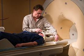
Magnetic Resonance Imagery, MRI, has revolutionized modern medicine, enabling to “see” inside the human body with no harm, contrary to most other methods such as X ray scanners for instance. MRIs are used everyday to diagnose tumours, sclerosis and oedemas.
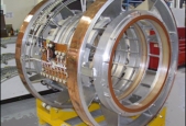
 MRIs use the small magnets inside the nuclei of the human body atoms to visualize what surrounds them (brain, muscles…). To do that, the nuclei magnets called “spin” must first be lined up thanks to a magnetic field in which the patient is put. The wider the magnetic field, the more accurate the image. To produce these fields, a strong electric current must flow through a coil of thousands of wire loops, called turns. If we used metal wires, such as copper for instance, they would heat so much they would melt. This is the reason why in all MRIs, the coil is made of a superconducting wire plunged in a very cold liquid such as helium.
There is no electric resistance, hence no heating. In addition to that, once the magnetic field has been created, the coil can be closed. The current (and hence the magnetic field) keeps flowing since there is no resistance.
MRIs use the small magnets inside the nuclei of the human body atoms to visualize what surrounds them (brain, muscles…). To do that, the nuclei magnets called “spin” must first be lined up thanks to a magnetic field in which the patient is put. The wider the magnetic field, the more accurate the image. To produce these fields, a strong electric current must flow through a coil of thousands of wire loops, called turns. If we used metal wires, such as copper for instance, they would heat so much they would melt. This is the reason why in all MRIs, the coil is made of a superconducting wire plunged in a very cold liquid such as helium.
There is no electric resistance, hence no heating. In addition to that, once the magnetic field has been created, the coil can be closed. The current (and hence the magnetic field) keeps flowing since there is no resistance.
Many researchers try to widen this magnetic field in order to see smaller details and get more accurate images, and hence better diagnoses.
 MRI is also used in chemistry, in biology and in physics to characterize matter, from the molecule to the solid. This is called NMR for “Nuclear Magnetic Resonance”.
In that case again, superconductors make these measurements possible by producing magnetic fields up to 500 000 times stronger than the earth magnetic field!