Three-dimensional imaging by single molecule localization (SMLM) allows resolutions of a few tens of nanometers, making this technique perfectly suited to the study of the arrangements of different proteins in the cell sample. However, fluorescent probe localization strategies still encounter certain limitations related in particular to a non-uniform axial precision or a depth of observation often limited to the first micron of the sample.
During the PhD’s work of Pierre Jouchet a new approach was developed for the localization of single molecules, called ModLoc, to address these various limitations. This new localization principle is based on the modulation of the fluorescence signal of single molecules through the use of a structured and time-modulated excitation, typically a sliding fringe array. The demodulation of this signal then makes it possible to trace the position of the molecule within the excitation pattern with nanometric precision. In theory the performance of this strategy showing a gain in precision of a factor of 3 compared to conventional localization methods, and we demonstrated an improvement of 2.8 along a lateral direction. Due to the random temporal nature of the emission of fluorescent probes in SMLM imaging, emission properties in dSTORM and DNA PAINT imaging implies to use frequency of demodulation faster than acquisition rate provided by the camera. We thus developed dedicated demodulation strategies based on fast optical element to bypass the acquisition frequency limits imposed by the camera.
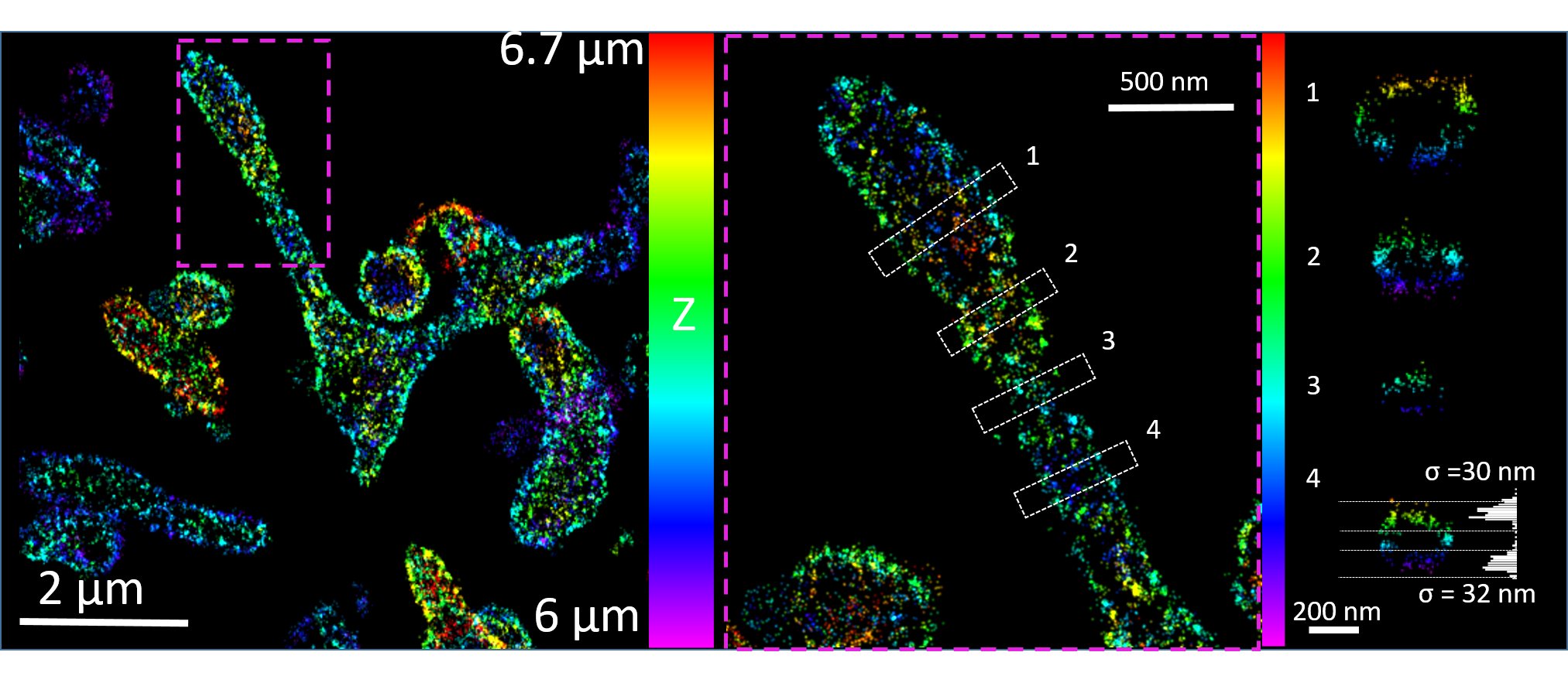
This modulation/demodulation approach can be applied to any direction to retrieve enhance precision. As axial localization of fluorescent molecules currently remains a challenge in single molecule imaging, we applied ModLoc to enhance this direction. The results obtained show a uniform localization accuracy of 6.8 nm and up to 7 microns in depth on both calibration and biological samples as represented below for the mitochondria network in COS7 cell located at 6 microns inside the cell. The robustness of the method for in-depth SMLM imaging is also demonstrated thanks to acquisitions made at a depth of 30 µm in aberrant media, as this type of information is generally not accessible in 3D SMLM imaging.
This work have been persented in various conferences (QBI 2019, FOM2019, SMLM2019, Photonics west 2020, BSP 2020) and you can find a detailed decription to extract axial information in Biorxiv .”Nanometric axial localization of single fluorescent molecules with modulated excitation” Pierre Jouchet, Clément Cabriel, Nicolas Bourg, Marion Bardou, Christian Poüs, Emmanuel Fort, Sandrine Lévêque-Fort. This work is now published in Nature Photonics, and here is a free reading link